What’s the diagnosis?
Plaster-like keratoses on the lower limbs


Multiple actinic keratoses usually appear in the setting of chronic sun damage and have an irregular, angulated and poorly defined shape with an irregular keratotic surface. Skin biopsy shows a disorganised keratinocyte architecture with nuclear atypia and an immature stratum corneum (parakeratosis).
Multiple plane warts are not usually keratotic and are often grouped. Skin biopsy shows vacuolated keratinocytes (koilocytes) reflecting viral cytopathic changes.
Stucco keratoses is the correct diagnosis and may be seen in up to 5% of elderly individuals. Originally described in Australia,1 the condition can be recognised as a clinically distinct phenomenon. Similar lesions have been described in tar workers. Stucco keratoses may be related to seborrhoeic keratoses as both show papillomatous epidermal hyperplasia and are not premalignant. Recently, mutations detected in stucco keratoses have further linked these as a possible variant of seborrhoeic keratoses. Individual lesions may be picked off without bleeding but tend to recur. Local treatment with keratolytic agents such as retinoic acid, glycolic acid, lactic acid or urea-containing creams may be helpful.
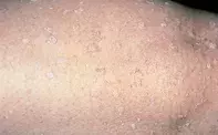
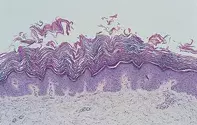
A 55-year-old woman presented with an eight-year history of numerous grey to white dry keratoses measuring 1 to 5 mm in diameter. The lesions were concentrated on her lower limbs (Figure 1) but were also present on both arms. The individual lesions were round to oval and were well demarcated. A skin biopsy showed a papillomatous epidermis with a loose and thickened basket-weave keratin layer (Figure 2). There were no viral inclusions or cytological atypia present.

