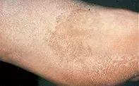
What’s the diagnosis?
Enlarging pigmented lesion on the foot


Café au lait macules may have an identical colour to this man’s lesion, but they appear in childhood, have an even colour rather than a mottled pattern, and are usually not on the sole. Biopsy shows epidermal hyperpigmentation.
Pigmented epidermal naevi also usually have their onset in childhood and have a more whorled or linear appearance. Biopsy often shows verrucose epidermal hyperplasia.
Chemicals such as self-tanning lotions may produce discolouration of the skin, but usually this is short lived and lacks the history of progressive growth.
Malignant melanoma of the sole is an important differential diagnosis. Malignant melanoma usually has a more variegated colour spectrum and dermoscopy shows a broad ridge pattern of pigmentation. Skin biopsy will help confirm the diagnosis prior to surgery.
Tinea nigra is the correct diagnosis in this case. It is caused by the presence of a superficial mould called Hortaea (Exophiala) werneckii. The mould is able to synthesise melanin and masquerade as a melanoma. The infection is usually localised to either a palm or sole. The diagnosis can be confirmed by skin scraping and culture or by skin biopsy. The mould will respond to topical imidazole creams, such as 2% miconazole or 2% ketoconazole, and also to keratolytic agents such as urea or Whitfield ointment (benzoic acid, salicylic acid).
Over a nine-month period, a 42-year-old man developed an enlarging, mottled, tan-coloured lesion on the sole of his left foot (Figure 1). A skin biopsy showed multiple pigmented spores and hyphal elements within the keratin layer (Figure 2).

