What’s the diagnosis?
Annular scaling lesions on the limbs


Tinea may occasionally present as widespread annular lesions. Clues to the diagnosis are pustules, and in large lesions there are often multiple inner rings. Biopsy or culture of the peripheral scale can be used to confirm the presence of fungi.
Pustular psoriasis is a rare condition which may appear as annular scaling and erythematous rings. The presence of numerous small pustules in early lesions, as well as skin biopsy demonstrating superficial epidermal pustules, may help in diagnosis.
Necrolytic migratory erythema is a rare disorder which produces multiple erythematous and scaling rings on the lower limbs, flexures and abdomen. Glossitis and diarrhoea may be seen. Skin biopsy shows a peculiar vacuolated superficial necrosis of the epidermis. An underlying islet cell tumour of the pancreas producing excessive glucagon is associated with this syndrome.
Subacute lupus erythematosus (SCLE) is the correct diagnosis in this case based on the clinical presentation and biopsy findings. In this patient ANA was negative but antibody to extractable nuclear antigens (ENA) was positive for Ro (SS-A) antibodies. SCLE has a lesser association with nephritis and neurological disease than acute systemic lupus erythematosus. The presence of Sjögren’s syndrome is a clue to more severe disease. Drug-induced SCLE has been described in patients taking a variety of drugs including terbinafine, thiazide diuretics, calcium channel blockers or ACE inhibitors. Sun avoidance and sunscreens are important general measures. Topical corticosteroid creams are also useful. The patient’s rash responded to hydroxychloroquine 200 mg daily.
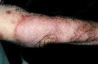
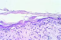
Over a three-month period, a 75-year-old woman developed multiple large annular lesions with scaling margins on the extensor aspect of her limbs (Figure 1). There was a longstanding history of arthritis of the right knee, which was treated with ketoprofen. Skin biopsy showed an atrophic epidermis with extensive vacuolar degeneration of the basal keratinocytes associated with a lymphocytic reaction (Figure 2).

