What’s the diagnosis?
An acute pustulovesicular eruption


Erythema multiforme may have an acute onset of erythematous and bullous lesions on the upper limbs, but it lacks a pustular element. The skin biopsy shows epidermal necrosis associated with an intensive lymphocytic reaction.
Halogenoderma may present with acute pustulovesicular lesions with a very similar morphology and is due to ingested halides, such as potassium iodide. Skin biopsy demonstrates pseudoepitheliomatous hyperplasia with neutrophil and eosinophil microabscesses.
Varicella may develop a pustular element and can be complicated by impetigo. In varicella lesions the vesicles are centrally placed, pustules are late, and the lesions are asynchronous. Bacterial and viral cultures and a skin biopsy showing viral cytopathic changes may be needed for diagnosis.
Sweet’s syndrome (acute neutrophilic dermatosis) is the correct diagnosis and belongs to a group of skin disorders characterised by sterile neutrophilic tissue reactions. Lesions are usually confined to the skin, but may rarely develop in the lungs, liver, kidney, bones or central nervous system. Major associated diseases are leukaemias, solid tumours, inflammatory bowel disease, connective tissue disease and respiratory infections. The lesions may spontaneously remit, but often recur. In severe or relapsing cases, oral prednisone is usually effective. Alternative treatments include potassium iodide, colchicine, dapsone or indomethacin. Lesions similar to Sweet’s syndrome can be induced by recombinant granulocyte colony stimulating factor.
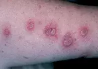
A 48-year-old woman presented with a five-day history of multiple, tender annular and arciform pustulo-vesicular lesions localised on her upper limbs (Figure 1), neck and face. There were associated symptoms of low grade fever, malaise and arthralgia.
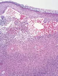
The skin biopsy revealed marked subepidermal oedema with an early blister. The underlying dermis showed sheets of neutrophils which had undergone leukocytoclasis (Figure 2). Vascular necrosis was not evident, although there were red cells in the subepidermal cavity.

